Dr. Patrick C. Nahirney (Associate Professor of Anatomy and Histology, Division of Medical Sciences, UVic) and Dr. William K. Ovalle (Professor Emeritus, Department of Cellular and Physiological Sciences [formerly Anatomy], Faculty of Medicine, UBC) recently released the third edition of their textbook/atlas Netter’s Essential Histology. Published by Elsevier, the revised textbook/atlas is used by healthcare students around the world, including UBC MD Undergraduate Program students across all four distributed sites in their first- and second-year histology course.
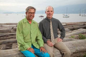
(L – R) Dr. William K. Ovalle and Dr. Patrick C. Nahirney
In addition to updated content focusing on normal cell ultrastructure and the microscopic anatomy of tissues and organ systems of the human body, Dr. Nahirney says the newest edition includes correlated histopathology to “keep pace with what’s been changing in the curriculum.”
The new text/atlas can also be accessed online via an enhanced digital eBook, which is available for a variety of devices. The eBook includes author-narrated video overviews of all 20 chapters and a virtual slide library of 20 high-resolution digitized light microscopic slides and 225 zoomifiable electron micrographs. There are also numerous “clinical points” that highlight relevant information on disease and cellular dysfunction.
According to the authors, the book is intended to be a valuable resource to both student learners and teachers by “awakening readers to both the intricacies of the human body and the sheer beauty of its cells, tissue, and organ systems.” It is also a beneficial tool for allied healthcare professionals, researchers, clinical residents, and medical laboratory technologists.
Companion to the book is an elegant set of Histology Flash Cards; for ease of use, the flashcards in the newest updated version (©2020) are cut-out cards.
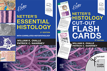
(L – R) Netter’s Essential Histology, 3rd edition, and Netter’s Histology Cut-out Flash Cards.
“The third edition is truly state-of-the-art and represents a great leap forward,” says Dr. Ovalle. “As co-authors, Patrick and I are very pleased with the latest edition.”
“I am also especially delighted that many of the electron micrographs that appear in our book were graciously donated to us by Dr. Bruce Crawford. He was a very dear colleague, mentor, and friend of ours over the years.”
The text/atlas also features drawings by renowned medical illustrator Dr. Frank H. Netter and other artwork by Drs. James A. Perkins, John A. Craig, Carlos A.G. Machado, and Joe Chovan.
Previously published in 2008 and 2013, Netter’s Essential Histology has been translated into Portuguese, Turkish, Korean, Greek, and Italian. A first South East Asia edition was also released in English in 2015.
The British Medical Association awarded Netter’s Essential Histology with its Best Illustrated Book Award in 2008, and Mark Yoffe, MD, cited the text/atlas in his 2015 article “The 25 Best Medical Books of All Time” on Medical Media Review.