With spectacular swirls of red, green, and blue, it’s easy to forget you’re looking at a precise image of a neuron – one of roughly 100 billion in the human brain.
The image was originally captured for a scientific study. But more recently, it’s been lauded for its stunning visuals. In February, it won the People’s Choice award from NeuroArt, a website powered by MBF Bioscience that celebrates the brain’s beauty.
“It’s exciting to have been selected, especially for an award voted on by our peers,” said Ryan Heron, a graduate from UBC’s Island Medical Program (IMP).
Drs. Heron and Patrick Nahirney, an associate professor in UVic’s Division of Medical Sciences (DMSC), submitted the image earlier this year after capturing it with an advanced fluorescence microscope in 2014. At the time, Heron was a second-year medical student who’d teamed up with Nahirney and Dr. Brian Christie, professor in the DMSC, for the project, which was supported by the Summer Student Research Program (SSRP).
Heron wanted to study the impact of omega-3 fatty acids supplements on the growth of neurites, branch-like structures that grow from neurons and, via electrochemical signals, communicate with neighboring neurons.
For Heron, who studied cellular biology as an undergraduate student, it was an opportunity to tie together his medical interests with his research acumen.
“This project was a highlight of medical school – the DMSC, its researchers, and its equipment are all top-notch,” he said. “I hope that more students take advantage of what’s available here.”
Dr. Heron graduated from the IMP in 2017. He then took a year off to support the IMP, working as a lab teaching assistant for Dr. Nahirney. He also created online learning modules, and was part of the team that developed the First Patient Program. This July, Dr. Heron will begin his family medicine residency in Edmonton.
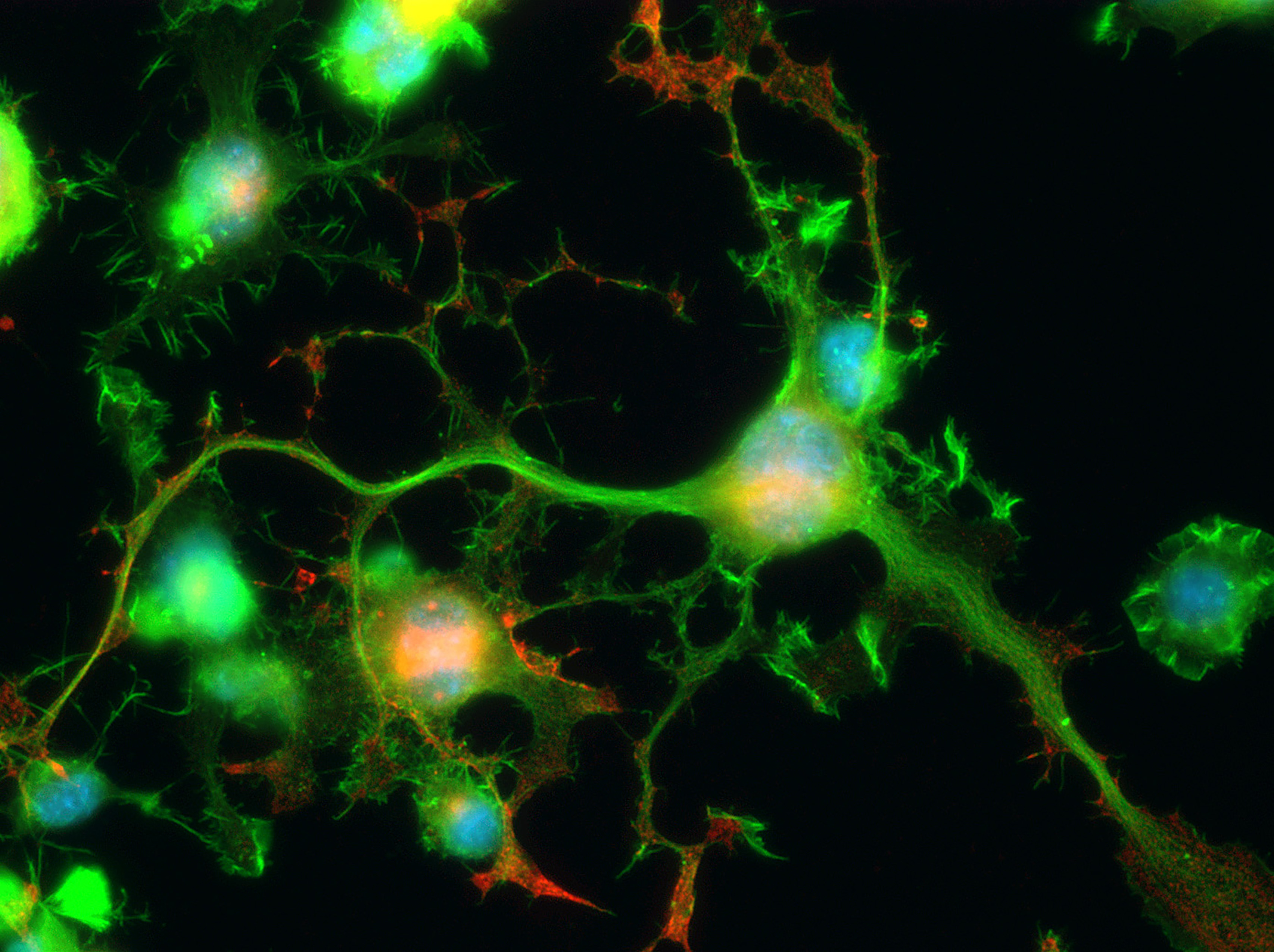
Photo: Drs. Heron and Nahirney captured their award-winning image using immunofluorescence microscopy, which uses antibodies, labelled with a specific fluorescent substances that emit different wavelengths of light, to better see the components of a cell – in this case, the nuclei (blue), tubulin (green), and synaptic vesicles (red).
Synaptic vesicles are made in the cell body, then travel along the tubulin cytoskeletal railway to the ends of the neuron’s outgrowth, where they serve a key role in the communication between neurons. The largest neuron (center-right) uses an elaborate network of outgrowths – neurites, which mature to become axons and dendrites – to connect with the neurites of other neurons.